app_localeMedicare honlap
Nyelvválasztás megjegyzése.
1 év
first-party



Ultrasound (US) is a gentle, painless procedure for detecting medical, endocrinological, urological, gynaecological, orthopaedic, and vascular problems.
Ultrasound is a range of sound waves that is not detected by the human ear. If the ultrasound waves are directed at the body by a probe, they are able to form an image, even while in motion, by means of the waves reflected from there.
Our colleagues are happy to help!
Our colleagues are happy to answer every question!
Do not eat for 6 hours before the abdominal and pelvic examination, only non-carbonated water is recommended for fluid intake. In case of pelvic or specifically bladder ultrasound, however, it is important that the patient arrives with a full bladder, so it is not advisable to urinate before the examination.
It is suitable for the examination of the abdominal and pelvic organs: liver, gallbladder, pancreas, spleen, kidneys, bladder, in women the uterus, ovaries, in men the testicles, the prostate can all be examined with ultrasound. With its help, an opinion can also be formed about the larger blood vessels running in the abdominal cavity, and the lymph nodes.
Organ size changes, structural abnormalities, developmental abnormalities can be seen with abdominal and pelvic US examination as well as cysts, stones, abscesses, benign fat and muscle tumours, malignant tumours, and abdominal fluid.
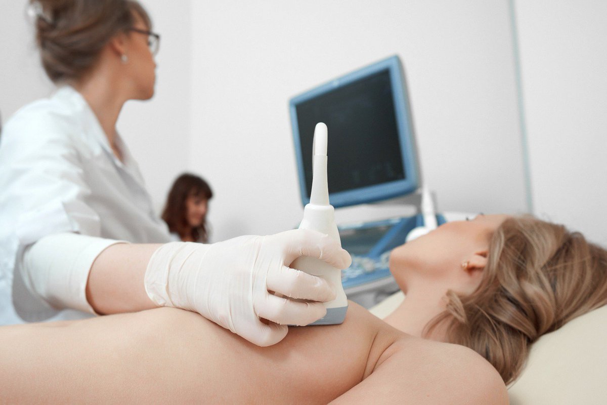
Ultrasound examination of the breasts is a painless examination without radiation exposure. The examination is suitable for detecting tissue and structural changes in the breasts (cyst, tumour, etc.).
The examination is performed while you are lying on your back and side. In addition to the breasts, the armpits are also examined.
Breast ultrasound does not require special preparation and can be performed during pregnancy.
Breast ultrasound is recommended for the detection of tissue and structural changes in the breasts (cysts, tumours, etc.) under the age of 35, while ultrasound, mammography and, where appropriate (in case of dense breast tissue), breast tomosynthesis are most effective methods for women over 35 years of age.
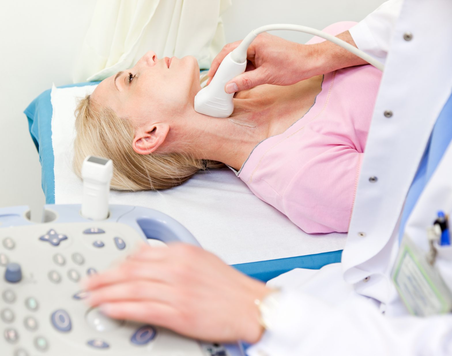
Ultrasound is an essential tool for the examination of the two lobes of the thyroid gland and the parathyroid gland. With its help, the structure and size of the lobes, nodules, cysts and inflammatory processes that may be present in the thyroid gland can be clearly seen.
In addition to the diagnosis of the thyroid and parathyroid glands, the thyroid gland US also plays an important role in the examination of the large salivary glands and cervical lymph nodes.
The structure of the carotid arteries and the blood flow can be judged well using a so-called Color Doppler, which is a coloured ultrasound examination.
This special ultrasound is excellent for the precise determination of the location, structure and size of vascular calcifications and the vessel stenosis they cause.
During the cervical soft tissue US examination, we examine the palpable nodules, superficial lesions and structural differences of the muscles on the neck and under the skin.
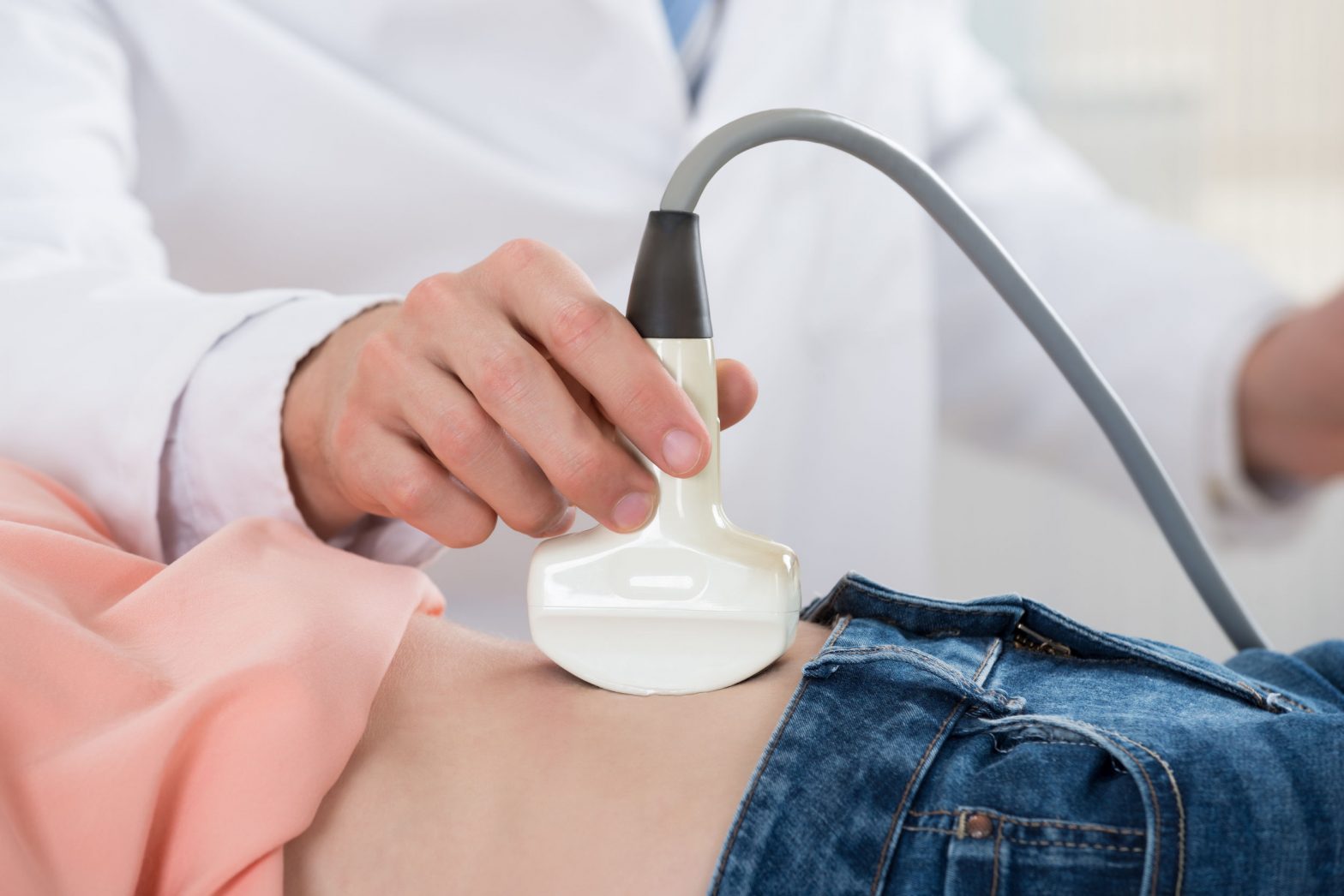
The primary goal of the pregnancy US is to screen for foetal abnormalities as soon as possible and with the greatest possible accuracy, as well as to shed light on the health of the mother.
Proof of pregnancy can be provided at the earliest in the second week after the absence of menstruation, if a pregnancy test performed at home shows a positive result.
Ultrasound is also used to examine the ligaments of the joints, tendons, fibres, cartilages, as well as the abnormal fluid deposits in the joints and the surrounding skeletal muscles. Joint ultrasound can also detect possible calcifications of the joint area, muscles, tendons, and arthritis
During the examination, the tissue structure of the testicles, the arteries and veins around the testis, and the layers of the scrotum and the epididymis are examined. Inflammations, tumours, and blood supply disorders can also be detected. Where appropriate, testicular hernia, testicular tumour, and varicose veins may be diagnosed.
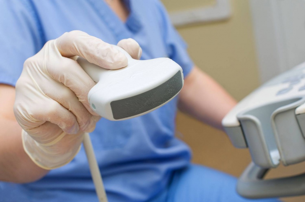
It is the examination of the subcutaneous soft tissues, muscles, tendons, adipose tissues, blood vessels, connective tissues, and lymph nodes. We also look at possible skin-related lesions.
Examination of the large vessels supplying the central nervous system as well as the large vessels running in the neck looks at the condition of the vessel wall and blood flow. Thanks to the examination, the predisposition to atherosclerosis leading to blood vessel stenosis or occlusion can be detected early.
Doppler ultrasound examines the circulation of different parts of the patient’s body and limbs. We check the function of blood vessels and any abnormalities that may exist. The examination can give an idea of venous inflammation, venous thrombosis, and venous valvular insufficiency.
Informational content only The textual content published on this page is for general information purposes only and does not constitute medical advice, a diagnosis, or a treatment recommendation. Proper assessment, examination, and therapeutic decisions always require a personal medical consultation and, where appropriate, further specialist opinion. Medicare Egészségközpont Zrt. makes every reasonable effort to keep the content up to date; however, it accepts no liability for the completeness or accuracy of the information published or for consequences arising from decisions based on it.