app_localeMedicare honlap
Nyelvválasztás megjegyzése.
1 év
first-party

A systemic infection, an infection that affects the whole body, can cause skin changes. Some skin lesions are hereditary (mole, freckles). Birthmarks can be the result of birth defects, while other skin lesions can be the result of an allergic reaction, poor circulation, or sensitivity to diabetes.
Our colleagues are happy to help!
Our colleagues are happy to answer every question!
A skin lesion is when a part of the skin looks different or shows abnormal growth. It can be divided into two major groups: primary and secondary lesions. Primary lesions appear at birth, such as moles. Secondary skin lesions develop after the irritation of the primary skin lesions. An infection, allergic reaction, or sensitiveness can also cause skin lesions.
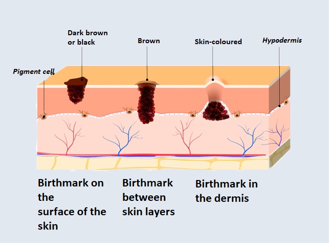
A sharp-lined lesion on the skin at birth or developing later is a birthmark. It is different in colour and surface from the skin. It is usually a few millimetres in size, but can grow much larger. Its colour is dark, yellowish brown, sometimes black. Its surface can be smooth or uneven, its shape may be circular or irregular.
In the case of later-formed moles, new ones may be formed as a result of UV radiation, in adolescence and pregnancy. Excision of a mole is necessary if it is aesthetically problematic, can be easily caught in anything due to its location and surface, or if a cancerous lesion (melanoma) is suspected.
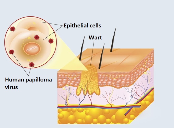
Warts are skin lesions caused by a viral infection. There are several different types that can form anywhere on the body: they most commonly appear on the fingers and back of the hands, the soles of the feet, the torso, and around the genitals. It can be caused by nearly 70 types of viruses, most of which are caused by a type of HPV (human papilloma virus).
Several types of warts can be distinguished: the most common is the common wart (verruca vulgaris), which is easily recognizable by its cauliflower-like appearance, the plantar wart (verruca plantaris), which due to its location can cause severe pain, the pool wart (mollusca contagiosa), which is bright, in the form of dented hemispherical outgrowths; and sexually transmitted genital warts (verruca genitalis).
The virus causing warts is most easily transmitted through damaged skin or in swimming pools with soaked skin. The risk of infection is particularly high in the case of a weakened immune system.
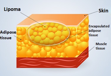
Lipoma (a lump of fat) is a benign skin tumour. The connective tissue lesion is well separated from the surrounding tissues. It is growing slowly. It mostly develops in the subcutaneous adipose tissue on the torso, legs or arms. The more connective tissue a lipoma contains, the harder it becomes.
Lipomas can occur alone isolated, or in small numbers. If they occur in larger numbers throughout the body, we can speak of lipomatosis. They mostly cause aesthetic problems. If a lipoma grows too large, numbness and pain may occur in that area, as this can put pressure on the nerves beneath the lipoma.
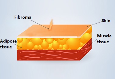
A fibroma is a soft, brownish lesion with a stem, which can occur in any part of the body. It is a benign cell proliferation of connective tissue cells. The cause of its development cannot be defined clearly, but both hormonal and genetic factors may play a role in its appearance. Mostly it is without complaint, but aesthetically it can be annoying.
In terms of its types, we distinguish between hard fibromas (low in cells), soft fibromas, also known as ‘game’, keloids (proliferation of scars due to injury or injuries), angiofibroma (there are dilated blood vessels among connective tissue cells) and cystic fibromas (there are lymphatic vessels among connective tissue cells).
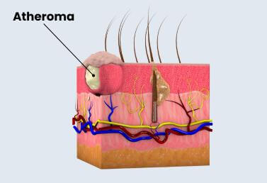
Atheroma, commonly known as a sebaceous cyst, is a benign sebaceous cyst that develops under the skin. It occurs when the duct of a sebaceous gland becomes blocked, preventing the sebum produced by the gland from draining out, but instead accumulates under the skin. This accumulated material has a thick, mushy consistency, hence the name.
The cyst has a connective tissue sheath that surrounds the sebum. The lesion is located directly under the skin and most often occurs on the face, scalp, neck, back, or trunk.
In terms of size, an atheroma can be a few millimeters or even several centimeters, especially if it persists for a long time or becomes inflamed. In most cases, it is between 1 and 5 cm in size.
Although atheroma is not malignant, it can become painful if inflamed or infected and may require medical intervention, such as surgical removal.
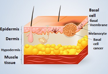
Basal-cell carcinoma is a slowly growing, malignant skin tumour. It develops from stem cells in the lower cell layer of the epidermis. It rarely causes metastasis. We distinguish between three types. Nodular basal-cell carcinoma is somewhat protruding from the skin, the edges are pearlescent, the centre is glossy, and turns into an ulcer over time. The superficial basal-cell carcinoma is sharp-edged, pale pink, extending towards the edges. Its middle may become small or scarred. Pigmented basal-cell carcinoma is an uneven, dark grey skin lesion.
The tumour usually develops on skin surfaces (face, ears, chest, back) that are permanently exposed to sunlight (UV-B rays). In some cases, however, it can also develop on areas of skin that are protected from sunlight. It often appears as a waxy tumour, but it can take different shapes.
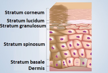
Skin colour is determined by a pigment (melanin) produced by specialized cells in the skin (melanocytes). The amount and type of melanin determines an individual’s skin colour.
Pigmentation disorder is a general term that includes hyperpigmentation disorders (increased staining) and hypopigmentation disorders (reduced staining). The most common diseases are melasma (hyperpigmentation) and vitiligo (hypopigmentation).
Some skin lesions can be malignant, therefore they need to be removed to avoid more serious illnesses. If the suspected cancer-prone lesion is not removed in time, its presence can later lead to a more severe stage or metastasis.
There may be other reasons for removing a particular lesion. Diagnostic causes, aesthetic appearance, or if it produces a symptom (e.g. pressure-sensitive or can be bumped) or inflamed or possibly infected.
Removal of each skin lesion requires different surgical techniques. The chosen technique depends on factors such as the severity and location of the lesion. The purpose of these surgeries is to completely remove the lesion and clean the area. A surgery takes about 15-45 minutes.
A successful skin lesion removal procedure effectively and completely removes any abnormal skin growth. In most cases, the removed lesion is sent to a laboratory and examined more thoroughly.
During the procedure, the surgeon excises the lesion and then sutures the wound so that the scar is as small as possible. Some patients’ skin heals abnormally. These patients may have larger-than-usual scars (keloid or hypertrophic scarring).
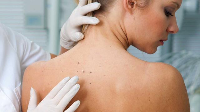
To diagnose the skin lesion, the specialist will perform a complete physical examination. The surgeon determines the type of lesion and considers the personal medical history and previous treatments.
It is very important that you continue to take care of your treated body and that you always keep it clean. A slight pink discoloration and pressure sensitivity around the edges of the wound is normal, but if it becomes redder or more painful over time, or if you develop a fever, make an appointment for a surgical check-up immediately.
The scar will initially be red and slightly raised but will usually lose its colour and size within a few months. A follow-up examination is recommended 7-14 days after surgery.
Informational content only The textual content published on this page is for general information purposes only and does not constitute medical advice, a diagnosis, or a treatment recommendation. Proper assessment, examination, and therapeutic decisions always require a personal medical consultation and, where appropriate, further specialist opinion. Medicare Egészségközpont Zrt. makes every reasonable effort to keep the content up to date; however, it accepts no liability for the completeness or accuracy of the information published or for consequences arising from decisions based on it.