app_localeMedicare honlap
Nyelvválasztás megjegyzése.
1 év
first-party

With the help of an elbow MRI examination, the doctor can see the state of the three bones meeting in the elbow, lesions following trauma, inflammation of the mucous tubes (bursa), the amount of fluid accumulating in the bursa, and diagnose tennis elbow.
With the help of an elbow MRI examination, the tendons, cartilage, the condition of the periosteum, the inflammations and the changes caused by them become visible to the doctor.
Our colleagues are happy to help!
Our colleagues are happy to answer every question!

Elbow MRI can diagnose several types of diseases. Your doctor may request an examination if you have the following complaints or illnesses.
Your doctor may consider an MRI scan of your elbow if you have long-standing local pain, reduced mobility and stiff limbs in the morning.
In all cases, it is recommended that your treating physician be consulted for details prior to the MRI examination, although the native examination does not require special preparation.
On the other hand, it is important to know that due to the strong magnetic field, it is forbidden to bring a device or object containing metal into the examination room! This applies to jewelry, piercings in the body, and some makeup products may also contain a substance that affects the success of the test.
Prior to an examination carried out with contrast agent, specialist consultation is essential. Kidney function laboratory results showing creatinine and carbamid values are also required to perform this type of examination. In the case of a contrast agent examination, you should not eat for 6 hours before the examination.
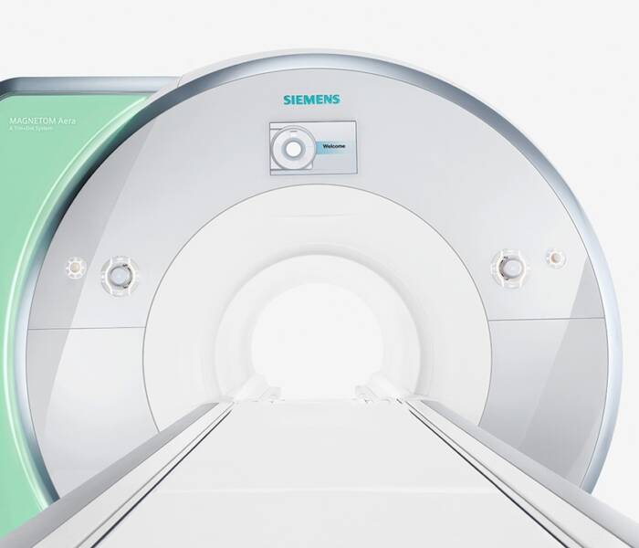
Following the pre-examination information, the operator will take you to the MR examiner room and then ask you to be seated on the examination bed. The operator adjusts the correct body position, places the coils required for the test, and then the test bed inserts you into the magnetic tube. It is important to remain still during the test, you can only change your body position on the instructions of the operator. The examination takes an average of 30 minutes, while you can communicate with our staff continuously through the built-in microphone and speaker. The machine makes a loud, rattling sound that can be reduced with ear protection.
The finding alone is not a diagnosis. After the elbow MR examination, please always consult a specialist with the result.
The contrast medium used for MR examination is gadolinium-based. In rare cases, hypersensitivity may occur, which may occur in the form of an allergic reaction.
The completed recording will be reviewed by a radiologist and the result will be available in our online system after 5 workdays.
Informational content only The textual content published on this page is for general information purposes only and does not constitute medical advice, a diagnosis, or a treatment recommendation. Proper assessment, examination, and therapeutic decisions always require a personal medical consultation and, where appropriate, further specialist opinion. Medicare Egészségközpont Zrt. makes every reasonable effort to keep the content up to date; however, it accepts no liability for the completeness or accuracy of the information published or for consequences arising from decisions based on it.